December 14, 2024
The Science Behind Breathing and Gas Exchange
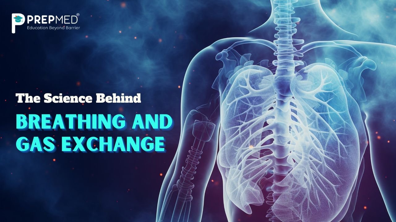
Breathing and gaseous exchange is one of the vital processes that is very important for every organism to sustain life. These processes help the body to get enough oxygen to function properly, necessary for respiration, and efficient removal of carbon dioxide which is a waste product of metabolism. The respiratory system comprises trachea, bronchi, bronchioles, alveoli, and lungs that help in the process of gaseous exchange.
When you inhale the air, oxygen reaches the lungs and goes down till alveoli which are described as tiny sacs present in the lungs that help in the exchange of oxygen and carbon dioxide during breathing. Oxygen enters the bloodstream by passing through the alveolar walls whereas carbon dioxide is transported from the blood into the alveoli and then excreted.
This process occurs due to the partial pressure of the gases and is important for regulating the process of homeostasis.
Let’s learn about the various respiratory organs of various organisms:
To understand the adaptations of various organisms, it is essential to understand the diversity of the respiratory mechanisms.
| Organisms |
Respiratory Organ |
| Terrestrial Mammals |
Lungs |
| Fish |
Gills |
| Amphibians |
Lungs and skin |
| Plants |
Stomata and Lenticels |
| Birds |
Air sacs and lungs |
| Insects |
Tracheal System |
| Earthworms |
Moist skin |
Understanding the Human Respiratory System
The human respiratory system is a detailed and complex network of various organs and structures that facilitates the process of gas exchange between the body and the environment. Respiration starts with the external pair of nostrils which leads to the inner nasal cavity.
Inside the nasal cavity, tiny specialized cells secretes mucus that helps in trapping dust particles. The nasal cavity leads to the pharynx and the pharynx has three different parts: oropharynx (passes food particles), nasopharynx (helps in airflow), and laryngopharynx.
Just below the nasopharynx, there is a structure called glottis which is surrounded by a cartilaginous and thin flap-like structure called epiglottis which helps in blocking the food particles from entering into the windpipe when you swallow the food.
Do you know?
- The larynx is also referred to as the voice box and is located at the top junction of the trachea and is essential for producing sound.
- It is held together with the support of thyroid and cricoid cartilage.
- The thyroid cartilage is more visible and prominent in males and it is most commonly known as the Adam’s apple.
- There are two vocal cords that are responsible for producing sound when air passes through them. The vocal cords are made up of mucous membranes.
- The trachea is the bridge joining the larynx and the lungs and is supported by C-shaped cartilaginous ring-like structures. This helps the trachea to remain open, thereby ensuring continuous airflow.
The gateway through the trachea
As you move down the trachea, the trachea divides into two bronchi, where each bronchi is responsible for passing the air into each lung. Inside these two lungs, the structures of bronchi further divide into smaller tune-like structures called bronchioles which are present throughout the lung.
The bronchioles then branch into other tiny ducts called alveolar ducts which lead to thin-walled tiny sacs of air called alveoli, which resemble bunches of grapes. An infundibulum is a structure that refers to a group of alveoli when clustered together, facilitating efficient gas exchange.
Let’s understand the mechanism behind respiration
Breathing is defined as the exchange of oxygen-rich atmospheric air between the lungs and the environment and the removal of carbon dioxide. This process occurs in two main phases: inspiration (expansion of the lungs) and expiration (contraction of the lungs).
When you inhale, during which the atmospheric air is drawn in, your diaphragm and the external and internal intercostal muscles between the ribs undergo contraction, which causes the chest cavity to expand. This causes the lung volume to increase, which in turn decreases the pressure inside the lungs than the pressure in the outside environment, that is, the pressure inside the lungs is lesser than the pressure in the atmosphere.
Alternatively when you exhale, during which the alveolar air is released out, your diaphragm and the external and internal intercostal muscles between the ribs undergo relaxation, which causes the chest cavity to shrink in its size. This causes the lung volume to decrease, which in turn increases the pressure inside the lungs than the pressure in the outside atmosphere, that is the pressure inside the lungs is more than the pressure in the atmosphere.
NOTE: If you see on an average, a healthy person normally breathes around 12-16 times every minute. The breathing rate can be easily calculated using a spirometer.
Get an idea about the respiratory volumes and their corresponding capacities
- Tidal Volume (TV): This is defined as the amount of air that is inhaled or exhaled during a regular respiration, under normal conditions. A healthy individual can inspire or expire around 6000-8000 ml of air every minute.
- Inspiratory Reserve Volume (IRV): This is defined as the additional amount of air that a person can inhale by a forceful inspiration. For a healthy person it typically ranges between 2500-3000 ml.
- Expiratory Reserve Volume (ERV): This is defined as the additional amount of air that a person can exhale by a forceful expiration. It is about 1000-1100 ml.
- Residual Volume (RV): The volume of air remaining back in the lungs after a forceful exhalation is known as the residual volume. It ranges between 1100-1200 ml.
- Inspiratory Capacity (IC): This is defined as the maximum volume of air a person can breathe in after a normal expiration. This can be calculated by taking a sum of the tidal volume (TV) and the inspiratory reserve volume (IRV).
- Expiratory Capacity (EC): This is defined as the maximum volume of air a person can breathe out after a normal inspiration. This can be calculated by taking a sum of the tidal volume (TV) and the expiratory reserve volume (ERV).
- Functional Residual Capacity (FRC): This is defined as the maximum amount of air remaining back in the lungs after a normal expiration. This can be calculated by taking a sum of the residual volume (RV)and the expiratory reserve volume (ERV).
- Vital Capacity (VC): The maximum amount of air a person can inspire after a forceful exhalation. This can be calculated by taking a sum of the tidal volume (TV) and the expiratory reserve volume (ERV) and inspiratory reserve volume (IRV).
- Total Lung Capacity (TLC): The maximum volume of air that the lungs can hold after a forceful inhalation. This can be calculated by taking a sum of the tidal volume (TV), expiratory reserve volume (ERV), residual volume (RV), and inspiratory reserve volume (IRV).
FAQS:
1. What are the three major layers of the diffusion membrane?
The diffusion membrane comprises three layers, the thin squamous epithelium of alveoli, the endothelium layer of the alveolar capillaries, and the basement substance in between these two layers.
2. What are the major organs of the respiratory system?
Nose, trachea, bronchi, bronchioles, lungs, nasal cavity, pharynx, and larynx are some of the major respiratory organs.
3. What are the major factors that affect the oxygen binding with hemoglobin?
The factors like partial pressure of carbon dioxide, hydrogen ion concentration, partial pressure of oxygen, and temperature affect the oxygen binding with hemoglobin.