January 04, 2025
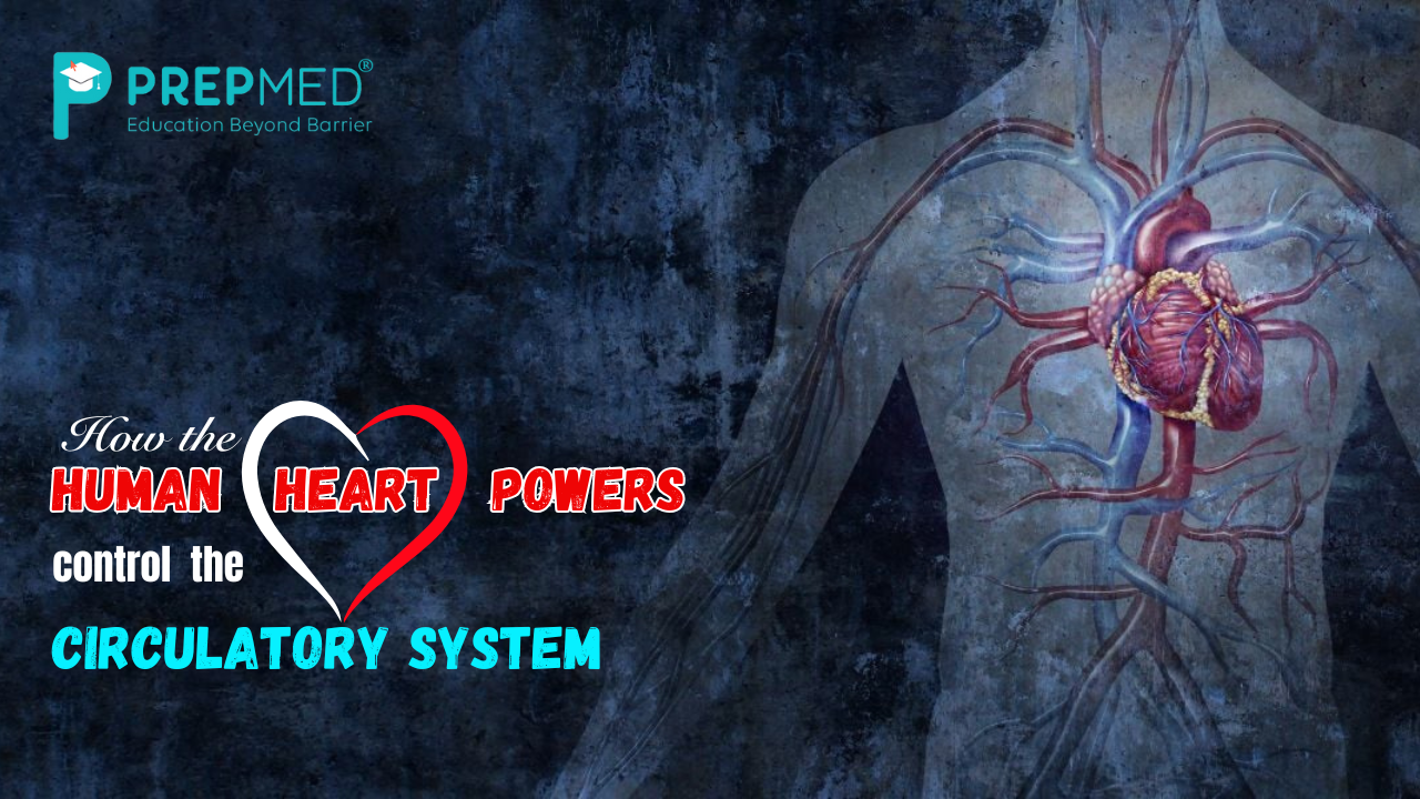
How the Human Heart Powers control the Circulatory System
One of the interesting topics taught in the NCERT Class 11 Biology book is the human circulatory system. This chapter focuses on the unique roles of the heart, blood vessels and blood through which nutrients, gases and waste products are transported in the body. The heart is a muscular organ that serves the function of a pumping of blood to all parts of the body through arteries, veins and capillaries. This system makes provision for the delivery of oxygen and nutrients to all parts of the cells and removal of carbon dioxide and other wastes.
The first area of concern of this chapter is the anatomy of the heart and its partitions, valves, and significant blood vessels. It describes how the pumping nature of the heart causes rhythmic contraction leading to the formation of a pressure gradient that transports blood. This article also discusses the distinctions of the major blood vessels as well as their roles in the human circulatory system. Furthermore, students are introduced to the components and uses of blood, functions of red blood cells, white blood cells, platelets, and plasma.
Thus, teaching and learning of the circulatory system enables students to acquire knowledge on how the body regulates itself by homeostasis and supports life. In this chapter, the reader is taken through a detailed analysis of the cardiovascular system with major focus on the relationship between the system and health and diseases.
Pathways of Circulation
The circulatory systems in animals can be broadly categorized into two types: let alone the open circulatory system, and the closed circulation system. In the open circulatory system, blood pumped by the heart passes into the open spaces through the large blood vessels into body cavities called sinuses. This type of circulation is best exemplified in molluscs and arthropods.
While, from mammals to fish, birds, reptiles and all other higher forms of animals possess a closed circulation system in which the blood has to percolate through closed vessels like arteries, veins, and capillaries. This system involves the heart, which is a muscular pumping organ.
In fish, the heart typically consists of two chambers one atrium and one ventricle. Most amphibians have only three-chambered hearts, while crocodiles have two-chambered heart, birds, reptiles, and mammals including man have four-chambered hearts. There are four chambers two auricles and two ventricles which enables the complete separation of oxygenated blood and deoxygenated blood.
Human Circulatory System
The human cardiovascular system is made of the heart, blood vessels including the arteries, veins and capillaries and the blood. The heart is a mesodermal organ which is located in the thoracic cavity between the lungs. It is enclosed in the double-layered sac known as the pericardium, which is filled with the pericardial fluid. The heart consists of four chambers two atria and two ventricles. These chambers are separated by walls that may be called septa.
Valves within the heart have the responsibility to ensure that blood flows in the right direction. The tricuspid valve is responsible for guarding the opening between the right auricle and the right ventricle. On the other hand, the bicuspid valve or the mitral valve protects the opening between the left auricle and the left ventricle. The Inter-auricular septum separates the right and left auricles whereas the Inter-ventricular septum separates the right and left ventricles. The auricle and the ventricle of the same side are separated from each other by atrioventricular septum. Semilunar valves are located at the exits of the right and left ventricles into the pulmonary artery and aorta, respectively.
Cardiac muscles form the entire structure of the heart. The thickness of the walls of the ventricles is relatively greater than that of the atrial walls. Furthermore, there are restricted cardiac muscles known as nodal tissue that is scattered all over the heart. Trace of this tissue exists in the anterior right corner of the right atrium in a region referred to as sinoatrial node (SAN). The second collection of nodal tissue is found in the lower left corner of the right atrium, in the region of the atrioventricular septum called the atrioventricular node (AVN). From the AVN, a collection of nodal fibres, known as the atrioventricular bundle (AV bundle) passes through the atrioventricular septae, in the top of the interventricular septum to branch into those for the right and left sides. These branches extend into fine fibers up to the ventricular muscle tissue on either side and are referred to as the Purkinje fibers.
Describe the cardiac cycle
The cardiac cycle is the series of events that happens during each heartbeat, thereby invoking both electrical and mechanical processes. There are two main phases that occur systole and diastole.
During the process of diastole, the ventricles of the heart undergo relaxation allowing the blood to flow into them. The process of atrial systole follows this, where the left and right atria, also known as left and right auricles in which the auricles contract, thereby increasing the blood pressure in the auricles to push blood into the ventricles. The AV valves are open and the semilunar valves are closed in this stage.
Then comes the process of ventricular systole, in which both the left and right ventricles contract, thereby increasing the blood pressure to pump blood into the arteries. In this stage, the AV valves are closed and the semilunar valves are open in this stage.
At last, the cardiac diastole takes place when the heart undergoes relaxation in order to get filled with blood once again. In this stage, both the auricles and the ventricles relax simultaneously. Opening of the bicuspid or the mitral valve takes place when the pressure of the left ventricle decreases below the left auricular pressure, allowing the blood to fill in the left ventricle.
Similarly the opening of the tricuspid valve takes place when the pressure of the right ventricle decreases below the pressure of the right auricle, thereby allowing the blood to fill in the right ventricle. The entire cardiac cycle is regulated by the electrical impulses generated by the Sinoatrial node (SAN) and atrioventricular node (AVN), making sure that the heart beats in a synchronised way. Throughout the process of a cardiac cycle, the heart regulates a rhythmic contraction pattern, ensuring a regulated blood circulation throughout the body.
Electro Cardio Graph
ECG is an electrical profile of the heart muscles during a cardiac cycle. For a detailed assessment of the heart’s function, multiple leads are connected to the chest part of the body. Here it is proposed to describe only a standard ECG. Every single wave in the ECG is lettered from P to T, each letter corresponding to a certain electrical event of the heart.
- P-wave is the electrical stimulus (or depolarisation) of atrial muscle resulting in the contraction of both atria.
- The QRS complex corresponds to ventricular depolarisation, from which the contraction of the ventricles originates from.
- The contraction begins shortly after Q and denotes the commencement of the systole.
- The T wave depicts the period of repolarization in which the ventricles go from an excited state to its normal phase.
- The top of the T-wave represents the end of the systole and the end of the T-wave separates diastole from systole. By analyzing the number of QRS complexes per a defined time period, it is possible to identify the heart beat rate of a given individual. Because of the similarity of ECGs collected from different individuals for the same lead configuration, any variation from this shape means an abnormality or disease that may be present in the patient’s body. Therefore ECG is of great clinical importance.
What do you understand about the Double Circulation?
The double circulation refers to the process in which the blood passes through the heart twice during every complete cycle. This process ensures that the oxygenated and the deoxygenated blood are separated efficiently which is essential for maintaining the metabolic needs of the body. The two atria and the two ventricles facilitate the process of double circulation.
1. Systemic Circulation:
- The oxygenated blood enters the left auricle from the lungs through the pulmonary veins.
- Contraction of the left auricle takes place, thereby pushing the blood into the left ventricle.
- Contraction of the left ventricle takes place, allowing the oxygenated blood to move through the aorta into the arteries, ensuring proper delivery of oxygen and nutrients to the tissues all over the body.
- After collecting carbon dioxide and other excretory products and ensuring proper delivery of oxygen, the deoxygenated blood returns to the right auricle via the inferior and superior vena cava.
2. Pulmonary Circulation
- The deoxygenated blood enters the right auricle from the body via the vena cava.
- Contraction of the right auricle takes place, thereby pumping blood into the right ventricle.
- Contraction of the right ventricle takes place, allowing the pumping of the deoxygenated blood to the lungs via the pulmonary arteries.
- The blood releases carbon dioxide and collects oxygen in the lungs.
- The oxygenated blood returns back to the left auricle via the pulmonary veins and completes the cycle.
The process of regulation of the cardiac activity
Normal activities of the heart are intrinsic, that is auto regulated by specialised muscles (nodal tissues), thus the heart is called myogenic. There is a special neural centre located in the medulla oblongata for the regulation of cardiac actions through the ANS. Stimulation of sympathetic nerves (which is a constituent of ANS) can speed up the rate of heart beat, enhance the signal strength of ventricular contraction and therefore the cardiac output.
On the other hand, the parasympathetic neural signals which is another branch of ANS reduces the rate of heart beat; the speed of conduction of the action potential and thus the cardiac output. Adrenal medullary hormones can also produce an elevated cardiac output.
Diseases of the circulatory system
- High Blood Pressure: High blood pressure refers to the blood pressure that is higher than the standard range (120/80 mm of Hg). It causes various heart diseases, strokes, and even problems in lungs, kidneys, and brain.
- Coronary Artery Disease (CAD): Coronary Artery Disease is also known as atherosclerosis and it is a disease in which the blood vessels carrying blood to the heart muscle is affected. It is caused by the formation of calcareous deposits, fats, cholesterol, and fibrous tissue which tend to shorten the lumen of arteries.
- Angina Pectoris: Also known as Angina, basically, acute chest pain as a symptom shows up when there is insufficient oxygen getting to the heart tissues. Angina can affect both male and female, children and adults, but it most frequently affects middle-aged and elderly people. It happens because of situations that impact blood circulation.
- Heart Failure: Heart failure means the condition of the heart when it will not pump blood in sufficient quantity to meet the needs of the body. It is sometimes called congestive heart failure because one of the primary signs of this illness is congestion of the lungs. Heart failure is not the same as cardiac arrest, which means the heart has stopped beating, or a myocardial infarction, which is a medical emergency that occurs due to the sudden damage of the heart muscle due to the inadequate blood supply.


